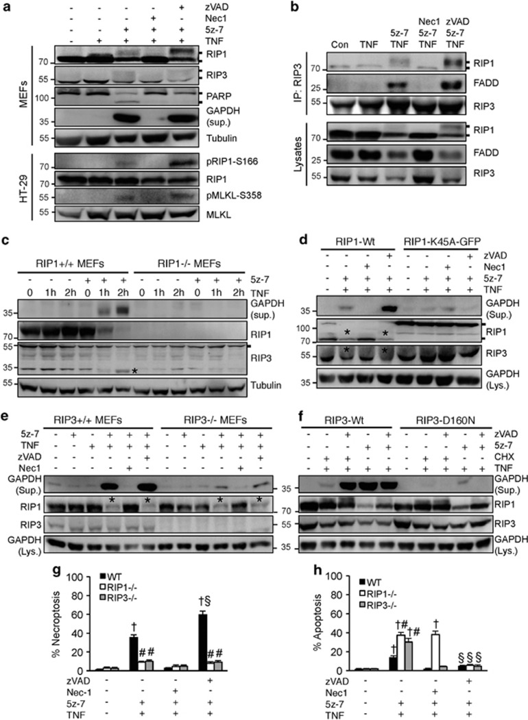
Figure 3.
Inhibition of TAK1 promotes RIP1 phosphorylation/activation and the RIP1–RIP3-FADD necroptotic complex formation. (a) Western blots for the indicated proteins from MEFs or HT-29 cells treated as indicated for 4 h. (b) Western blots for the indicated proteins following IP with an anti-RIP3 antibody from extracts of MEFs treated as indicated for 1 h. (c) Western blots with the indicated antibodies from cellular extracts of RIP1+/+ and RIP1-/-, MEFs treated with TNFα for 0, 1, and 2 h in the presence or absence of 5z-7. (d) Western blots with the indicated antibodies from cellular extracts of RIP1-/-, MEFs reconstituted with wild-type (Wt) or K45A mutant of RIP1, then treated as indicated for 4 h. (e) Western blots with the indicated antibodies from cellular extracts of RIP3+/+ and RIP3-/-, MEFs treated as indicated for 4 h. (f) Western blots with the indicated antibodies from cellular extracts of RIP3-/-, MEFs reconstituted with wild-type (Wt) or the D160N mutant of RIP3, then treated as indicated for 4 h. (g, h) Necroptosis (PI positive without chromatin condensation) and apoptosis (PI negative with chromatin condensation) in wild-type, RIP1-/-, and RIP3-/-, MEFs treated as indicated for 4 h. †P<0.05 versus Control; #P<0.05 versus WT; §P<0.05 versus 5z-7 plus TNF

