
Fig. 4.
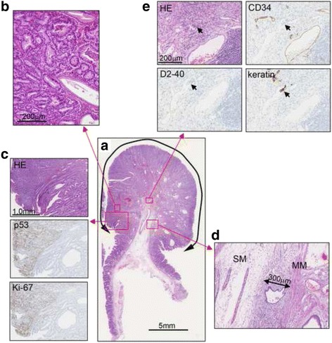
Histological findings. a. Hematoxylin and eosin (HE) staining. b. High magnification image of the boxed area. c. HE, p53, and Ki-67 stainings of the border between the hyperplastic glands and the tumor glands. The tumor lesions were strongly positive for p53 and Ki-67. d. Submucosal invasion in the stalk, with an invasion depth of 300 μm (MM, muscularis mucosae; SM, submucosal layer). e. HE, cluster of differentiation 34 (CD34), D2-40, and keratin staining. In CD34-negative and D2-40-positive lymph ducts, keratin-positive cells with acidophilic cytoplasm and large nuclei were observed, indicating lymphatic invasion (arrows)