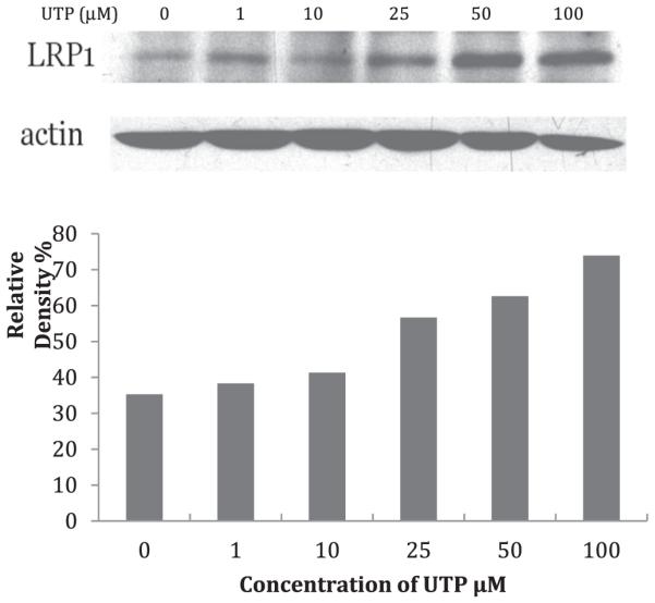
Fig. 1. Protein abundance of LRP1 expression in mouse VSMCs.

Serum-starved VSMCs were incubated in the absence or presence of UTP (1, 10, 25, 50, 100 μM). (Upper panel) Detection of LRP1 was performed using mouse anti-LRP1 monoclonal antibody (1:3000 dilution; Abcam, Cambridge, MA) followed by horseradish peroxidase-conjugated donkey anti-mouse polyclonal antibody (1:10,000 dilution Abcam, Cambridge, MA). For signal normalization, membranes were probed with polyclonal anti-β-actin antibody (1:2000 dilution). (Lower panel) The results show that LRP1 expression is increased in a UTP dose-dependent manner.
