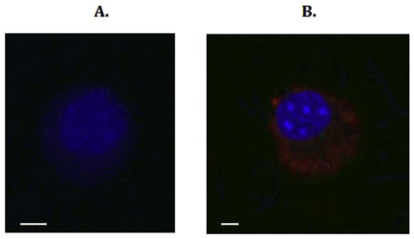
Fig. 4. UTP stimulates matrix-bound agLDL uptake in VSMCs expressing WT P2Y2R but not in VSMCs expressing a mutant P2Y2R that does not bind FLN-A.

VSMCs from P2Y2R−/− mice were transfected with (A) a mutant P2Y2R that does not bind FLN-A or (B) WT P2Y2R. Cells were grown on top of matrix, incubated with 50 μg/ml agLDL for 4 h at 37 ° C and then stimulated with 10 μM UTP. Lipids were stained by incubating cells with 1,1′ dioctadecyl-3,3,3′ ,3’-tetramethyl-indocarbocyanine (DiI; red) and cells were visualized using a Zeiss LSM 700 confocal microscope. The nuclei were stained by incubating cells with 1 μg/ml Hoeschst dye (blue). The images are representative of results from at least 3 independent experiments. Scale bar represents 50 μm. (For interpretation of the references to colour in this figure legend, the reader is referred to the web version of this article.)
