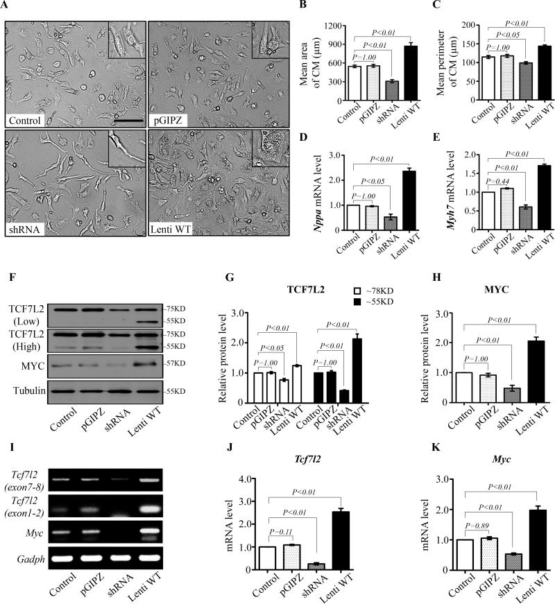
Figure 5.
TCF7L2 levels control c-Myc expression in cultured neonatal rat cardiomyocytes (NRCMs). A to C: Morphological changes of NRCMs by TCF7L2 transfection. Cells transferred with Tcf7l2 shRNA were narrow and thin spindle-like shape (A, left lower) with smaller size (B) and perimeter (C) compared with non-infected control group. NRCMs enlarged and varied from round-spindle to polygonal by transfection with short form full-length Tcf7l2 (Lenti WT) (A, right lower), while the size and perimeter of cells dramatically increased (B and C). D and E, Nppa and Myh7 mRNA levels were downregulated by Tcf7l2 shRNA, but upregulated by infection with full-length Tcf7l2. F to H: Representative TCF7L2 and c-Myc Western blots (F) as well as fold changes of TCF7L2 (G) and c-Myc (H) in NRCMs transfected with no virus, control virus (pGPIZ), Tcf7l2 shRNA, and full-length short form Tcf7l2. I to K: Semi-quantitative RT-PCR (I) and fold increases by real-time RT-PCR of Tcf7l2 (J) and c-Myc (K) in NRCMs transfected with no virus, control virus (pGPIZ), Tcf7l2 shRNA, and full-length short form Tcf7l2. Data are representative of five independent repeats. P<0.05 indicated statistically significant differences compared to control groups. Scale bar, 100μm.

