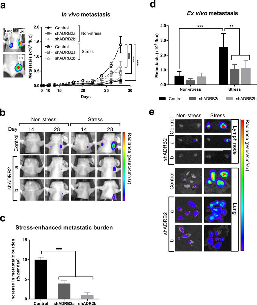
Fig. 4. β2AR knockdown in MDA-MB-231HM breast cancer cells impairs metastasis in vivo.
(a) Left panel: Representative in vivo bioluminescence image of the orthotopic MDAMB-231 breast cancer model showing primary tumor (PT) and spontaneously disseminated metastasis in lymph node (LN) and lung 28 days after tumor cell injection. Right panel: Quantification of distant metastasis by bioluminescence imaging in mice with tumors derived from scramble control or β2AR-deficient cells (shADRB2a and shADRB2b). Mice were exposed to non-stress or stress conditions. (n = 5 at each time point). (b) Representative images of metastasis in vivo at day 14 and 28 after tumor cell injection. Note: the primary tumor is not visible. Scale: min. 3 × 10⁶ photons/sec; max. 1 × 10⁸ photons/sec. (c) The magnitude of the effect of stress on metastatic burden was computed as the difference in the rate of metastatic progression under non-stress and stress conditions. (d) Ex vivo quantification of distant metastasis in target organs that were harvested from mice 28 days after tumor cell injection (n = 5). (e) Representative images of metastasis in lung and lymph node ex vivo at 28 days post tumor cell inoculation. Scale: lymph node: min. 5 × 10⁶; max. 1 × 10⁸ and lung: min. 2 × 10⁵; max. 8 × 10⁶ photons/sec. Data represent mean ± standard error. *p < 0.05 and ***p < 0.001 by (c) one-way ANOVA or two-way ANOVA (a) with or (d) without repeated measures followed by post hoc Tukey’s planned comparison tests.

