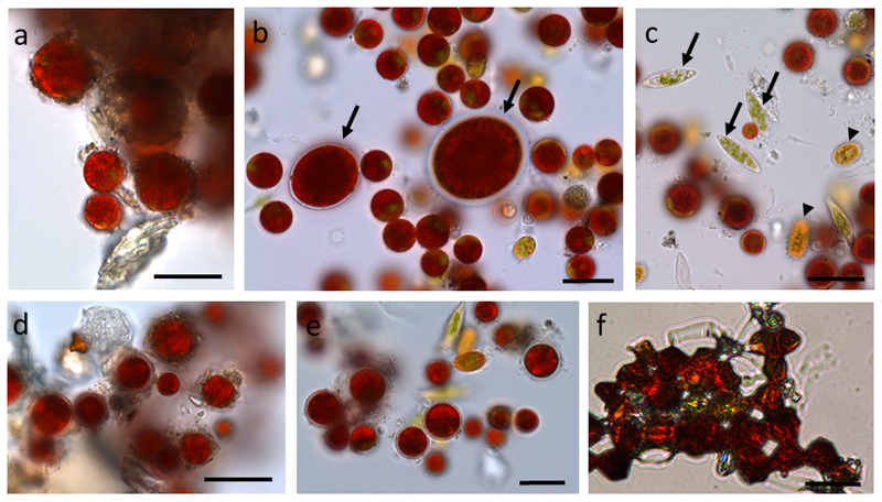
Fig. 1.
Light microscopy of snow algal cells from different field samples (S1, a, d) and (WP79 b-c, e-f). (a) Chlamydomonas nivalis covered with debris, (b) Chlamydomonas nivalis, Chlainonmonas sp. cells are marked with an arrow, (c) Chloromonas rosae var. psychrophila (arrows), Chloromonas brevispina (arrowheads), (d) sample S1 plasmolysed in 1,600 mM sorbitol, (e) sample WP79 plasmolysed in 1,600 mM sorbitol, (f) sample WP79 desiccated. Bars 20 µm

