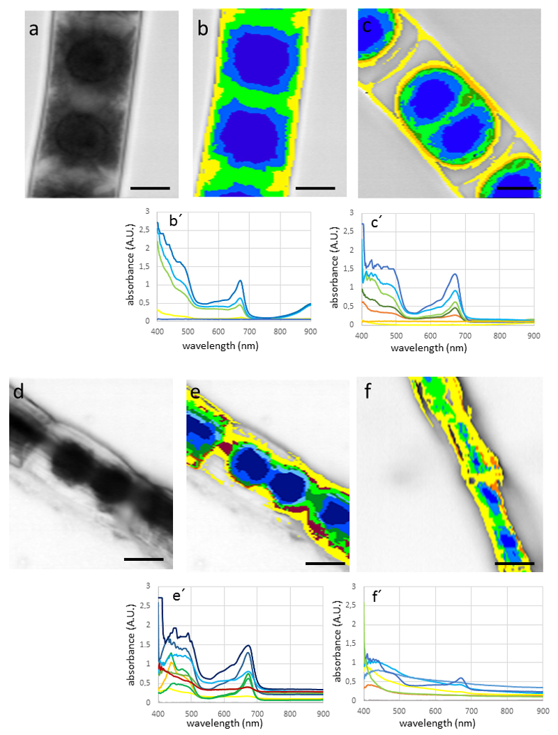
Fig. 5.
Hypersectral images and corresponding spectra (below) of Zygnema sp. (A-E) and Zygogonium ericetorum (F) cells. (a) bright field image, (b) hyperspectral image of the same cell, (c) cells plasmoylsed in 800 mM sorbitol, retraction of the protoplast, (d) bright field image of air dried cells, (e) corresponding hyperspectral image, (f) desiccated cell of Zygogonium ericetorum, note the irregular shrinkage of the cells. Bars 10 µm

