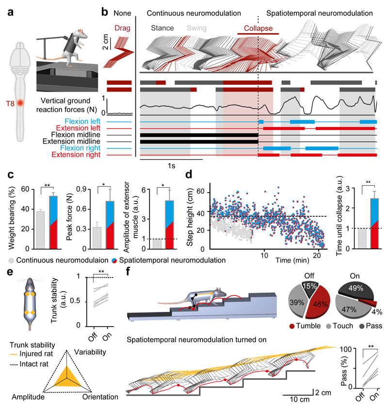
Figure 6. Spatiotemporal neuromodulation improves motor control after clinically relevant SCI.
(a) Rats received a contusion at T8. (b) Locomotion was recorded 14 days after injury on a treadmill. Continuous neuromodulation led to rapid fatigue, but spatiotemporal neuromodulation instantaneously restored locomotion. (c) Histogram plots reporting the maximum weight–bearing capacities, maximal vertical ground reaction forces, and amplitude of extensor muscle activity measured in the same rats during continuous versus spatiotemporal neuromodulation (paired t–test, n = 6, *, P < 0.05, **, P < 0.01). (d) Left, successive step heights measured during a sequence recorded in the same rat under both paradigms. The horizontal line indicates the mean of step height measured in intact rats (n = 3). The shaded area reports the SEM. Right, bar plots report the normalized duration of locomotion (paired t–test, n = 6, P < 0.01). (e) Rats were recorded during quadrupedal locomotion at two months post–SCI. Parameters related to trunk dynamics were computed to obtain an index quantifying trunk stability relative to intact rats. The improvement of this index is shown for each rat individually (paired t–test, n = 6, P < 0.01). (f) Stick diagram decomposition of hindlimb and trunk movements together with hindlimb endpoint trajectory during staircase climbing under spatiotemporal neuromodulation. The diagrams report the percent of tumbles, touches and passes over the steps under both conditions. The percent of successful passes over the step is shown for each rat individually (paired t–test, n = 6, P < 0.01).

