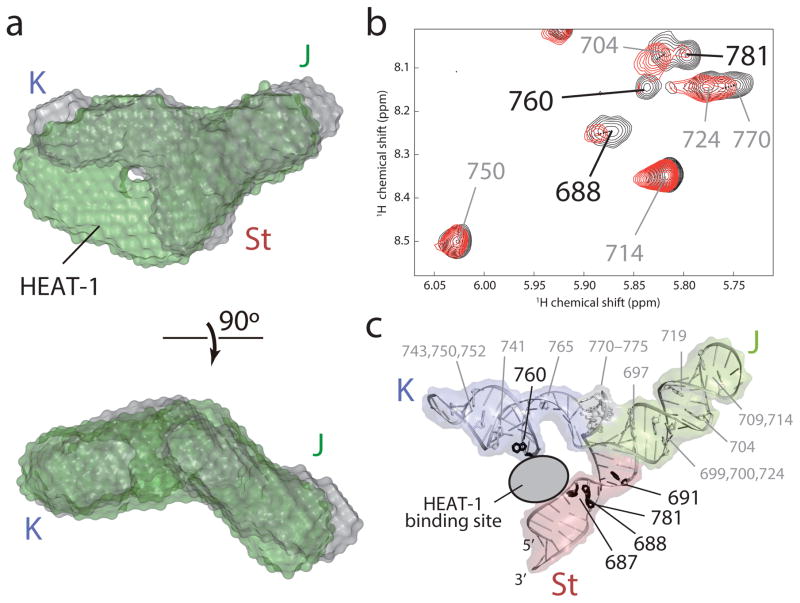
Figure 5. Interaction of the J-K region with the HEAT-1 domain of eIF4G.
(a) Overlay of the SAXS ab initio envelope structure of the J-K region (gray) with that in complex with the HEAT-1 domain (green). The top panel is shown from the same perspective as in Fig. 3a, left panel. (b) 1H-1H NOESY spectra of [u-2H, {H1′,H2′,H2,H8}-Ade] J-K region in the absence (black) and substoichiometric presence (red) of the HEAT-1 domain. Assignments of the intraresidual H1′–H8 NOE signals are labeled. Residues whose NOE signal intensities were substantially reduced by the addition of the HEAT-1 domain are labeled in black. (c) Mapping of the adenosine residues in (b) onto the structure of the J-K region. Adenosine residues whose signal intensities are substantially reduced upon addition of the HEAT-1 domain are colored black whereas the others are white.

