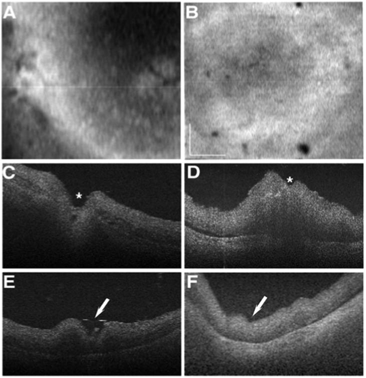
Figure 2.

Retinal disorganization of the affected donor retina. Ex-vivo SD-OCT en face imaging of the retina of affected donor (B) and age-matched control (A) control eye. C-F: SD-OCT B-scans of affected donor (D, F) and age-matched control (C, E) eye. Asterisks show optic nerve head, and white arrows show macula. Bar = 0.5 cm.
