
Figure 4.
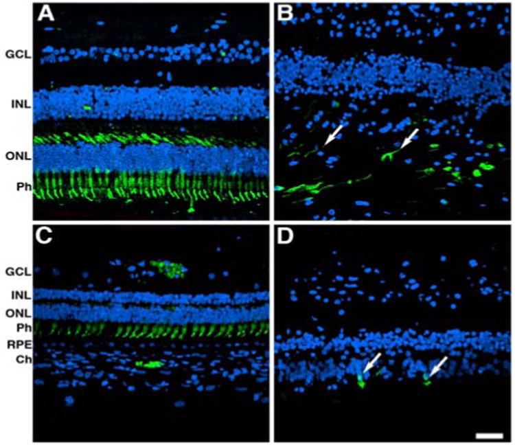
Significant alterations in the cones both in the perifovea and in the periphery of affected donor. Representative immunofluorescent staining of cone arrestin in control retina in the perifovea (A) and periphery (C) compared to affected donor retina in the perifovea (B) and periphery (D) were analyzed by confocal microscopy.
GCL: Ganglion Cell Layer; IPL: INL: Inner Nuclear Layer; ONL: Outer Nuclear Layer; Ph: Photoreceptors; RPE: Retinal Pigment Epithelium; Ch: Choroid. Bars =40 μ.