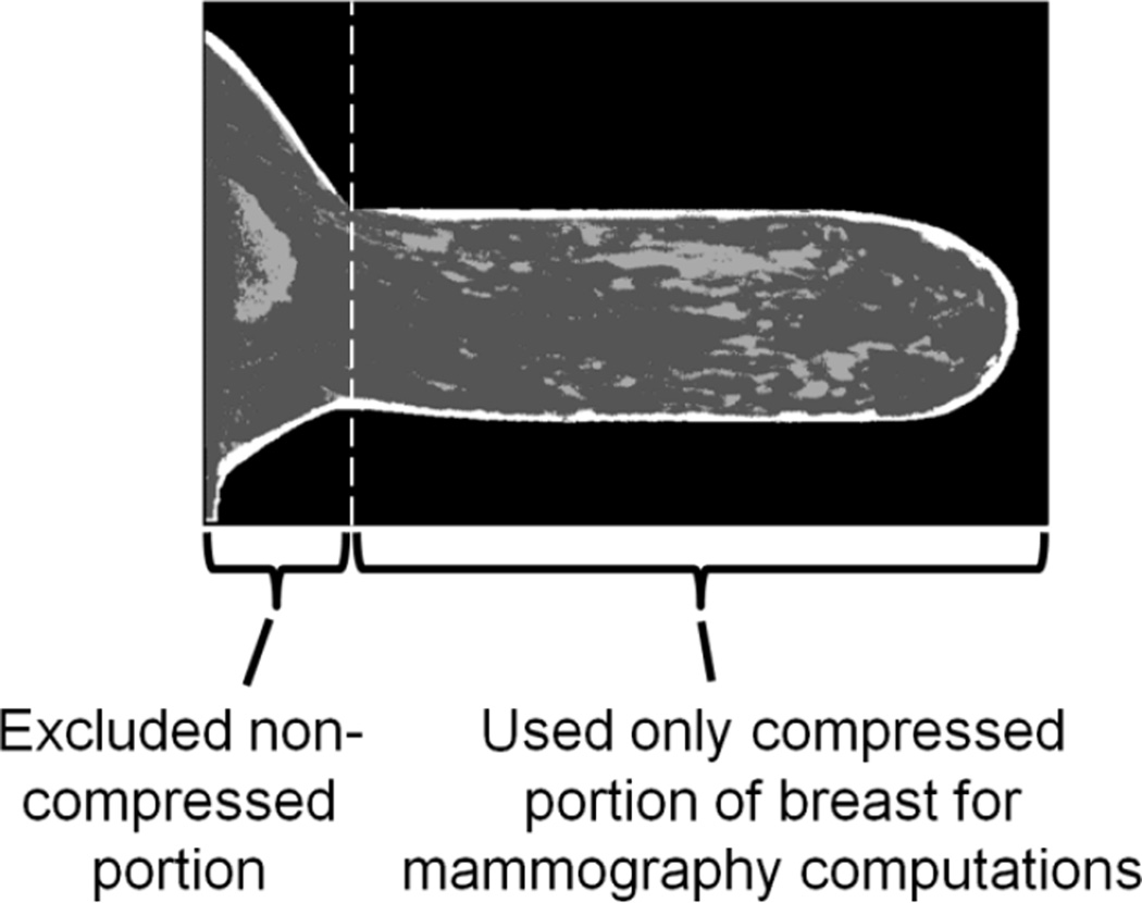
Figure 12.
Sagittal slice of one of the DBCT patient images that was classified and compressed to represent a breast with its real patient tissue structure undergoing mammography in the study by Sechopoulos et al (2012). Figure from Sechopoulos et al (2012). Copyright (c) 2012, American Association of Physicists in Medicine (AAPM).

