Figure 6.
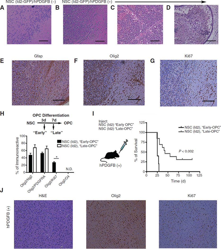
Histologic examination of glioma arising in hPDGFB (+) mice orthotopically inoculated with NSC (Id2-GFP). A and B, photomicrographs of hematoxylin and eosin (H&E) stained histologic sections showing the ventral forebrain of a WT animal (A) and an hPDGFB (+) age-matched littermate (B) orthotopically inoculated with NSC (Id2-GFP). Original magnification, ×4; scale bars, 200 µm. C and D, photomicrographs of H&E stained histologic sections of tumors obtained from hPDGFB (+) animals inoculated with NSC (Id2-GFP). Original magnification, ×20; scale bars, 100 µm. E–G, adjacent histologic sections to B immunostained for Gfap, Olig2, and Ki67 (brown) and counterstained with hematoxylin (blue); original magnification, ×4. H, quantification of Olig2/Ng2, Olig2/PDGFRa, and Olig2/Ki67 double-positive cells detected using immunostaining at 3 and 7 days following the initiation of OPC-directed differentiation. Data, mean ± SD from two independent experiments plated in triplicate; *, P < 0.05. I, Kaplan–Meier survival plot from NSC (Id2-GFP) inoculated 3 days after initiation of after OPC-directed differentiation compared with NSC (Id2-GFP) inoculated 7 days following differentiation. J, histologic sections taken from an hPDGFB (+) animal following inoculation NSC (Id2-GFP) differentiated for 3 days under OPC enrichment conditions. Sections are stained with H&E, Olig2, and Ki67 (brown). Original magnification, ×10; scale bars, 100 µm.

