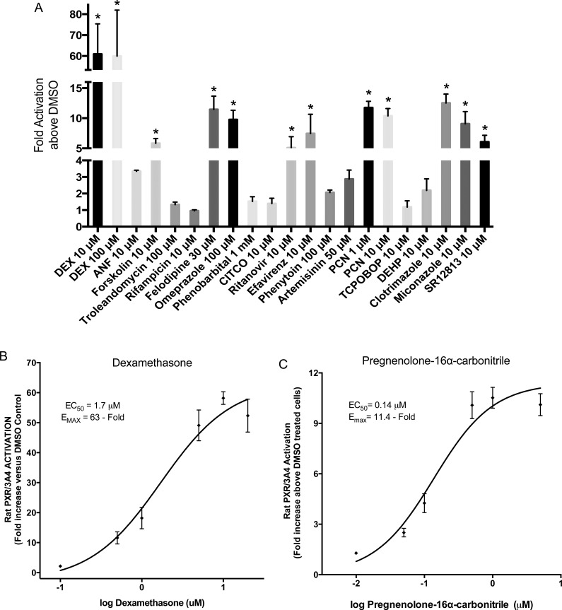
Fig 3. Rat PXR transactivation by 19 compounds in a stable cell line.
A rat PXR stable cell line over-expressing rat PXR and a luciferase reporter vector containing the CYP3A4 proximal and distal enhancer regions [14] was used to evaluate activation of rat PXR by various compounds (A). rPXR cells were seeded in 96-well plates and treated with the 19 compounds for 48 h before fluorescence (cell viability) and luminescence (transcriptional activation) detection. All luminescence values were normalized for cell viability. The data represent the mean ± SE from three independent experiments in triplicate expressed as fold activation above vehicle control treated cells. An asterisk denotes compounds exhibiting significant difference from their respective vehicle control at a level of p < 0.01. (C-D) Dose-response curves for two rat PXR positive controls, Dexamethasone (B) and Pregnenolone 16α- carbonitrile (C). Concentrations were 0.1, 0.5, 1, 5, 10 and 20 μM for Dexamethasone (B) and 0.01, 0.05, 0.1, 0.5, 1 and 5 μM for Pregnenolone 16α- carbonitrile (C). Results are expressed as fold activation above 0.1% DMSO treated cells. Data represents the mean ± SE from three independent experiments in triplicate. EC50 and EMAX values were calculated using nonlinear regression of typical log dose-response curves.

