
Figure 7. RA patient sera show strong antibody recognition of citrullinated R289 that is buried in structure of BiP.
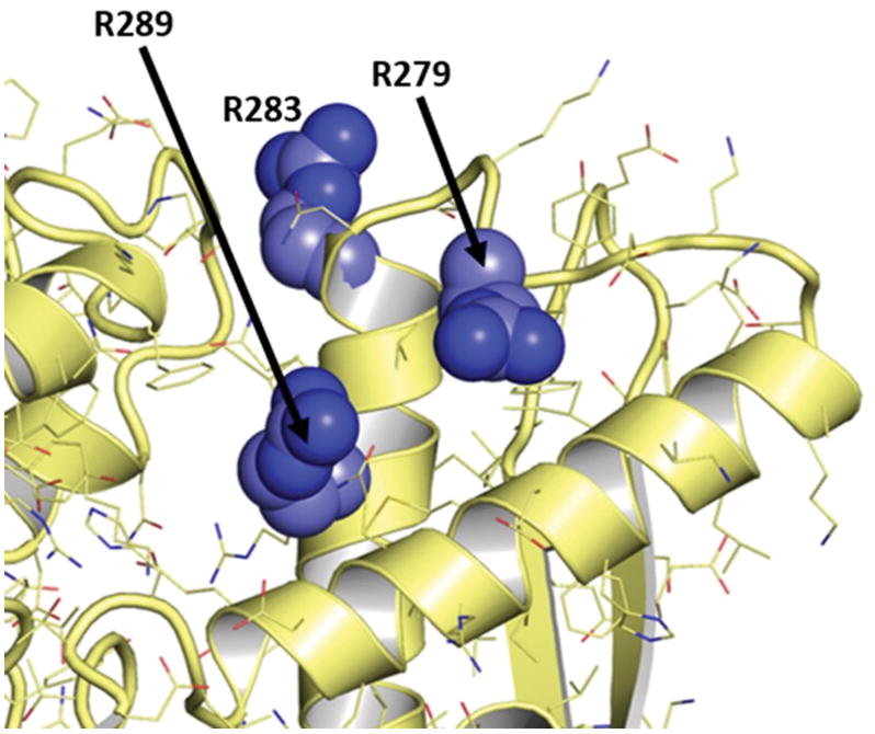
Nearby sites at R279 and R283 are solvent-exposed, but they are not targeted by autoantibodies derived from RA patient sera. Structure taken from PDB ID 3IUC (57).