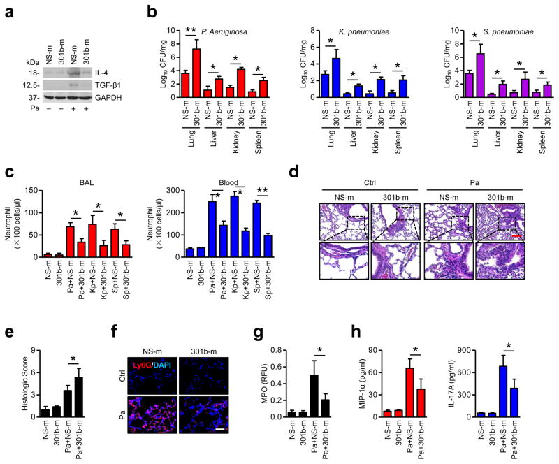
Figure 3. Enforced expression of miR-301b suppresses bacterium-induced anti-inflammatory responses in vivo.
(a) Mice (n=3) were i.v. injected with NS-m or 301b-m (50 μg/mouse, 24 h), then infected with Pa (1×10⁷ CFU), Kp (1×10⁵ CFU) or Sp (5×10⁶ CFU) for 24 h. Lungs from mice infected with Pa as above were collected and homogenized for immunoblotting for IL-4 and TGF-β1. (b) Organs from mice were collected and homogenized for CFU assay. (c) Neutrophils in BAL and blood were counted by H&E staining. (d, e) Lungs were embedded in formalin for H&E staining and lung injuries were double-blind scored. Scale bar=100 μm. Insets showing tissue injury and inflammatory influx. (f) Lungs were embedded in formalin for immunostaining with a Ly6G antibody to show neutrophil infiltration. Scale bar=50 μm. (g, h) After treatment as above, MPO and ELISA assays were performed to test myeloperoxidase activities in lung homogenates or MIP-1α and IL-17A levels in BAL, respectively. Panel a, d, e, f, representative from triplicate samples; b, c, g, h, data are from 3 mice/group, means+SD. *, p<0.05, **, p<0.01. One-way ANOVA with Tukey’s post hoc.

