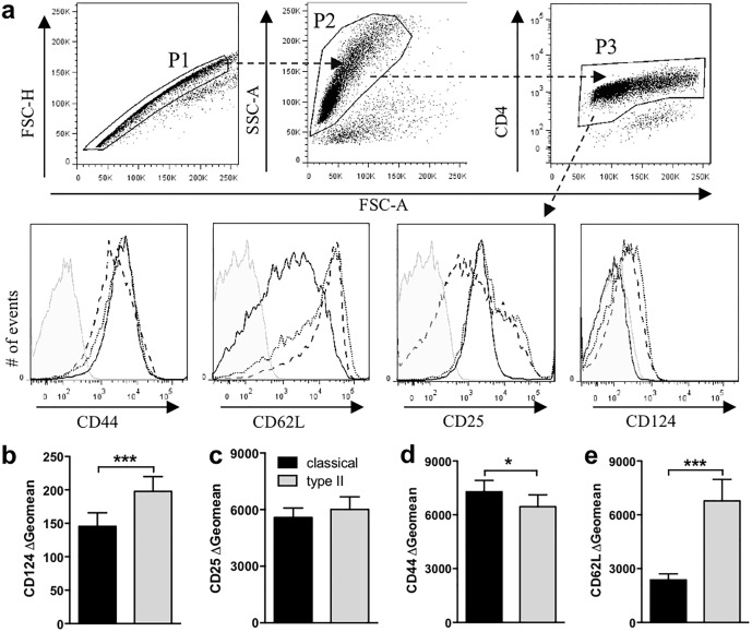
Fig 2. Type II macrophages reduced T cell activation markers.
Macrophages were primed with IFN-γ (20 U/ml) overnight. IFN-γ was removed with warm media and the macrophages were stimulated with or without LPS (200 ng/ml) in the presence or absence of IC (10 IC/macrophage). After four hours, purified CD4+ 2D2 T cells and MOG (25 μg/ml) were added and cultured for 72 hours. CD124 (b), CD25 (c), CD44 (d), and CD62L (e) expression was assessed by flow cytometry. (a) Gating strategy to assess cell surface markers on T cells (P1, single cells; P2 live cells by FSC vs SSC; P3 CD4+ cells). Grey filled = Isotype control; solid line = classical macrophage; dotted line = type II macrophage; dashed line = unstimulated macrophage. (b-e) T cells cultured with type II macrophages express lower levels of CD124 (b) and higher levels of CD62L (d) compared to those cultured with classical macrophage. Shown are the geometric means from 17–19 individual experiments. *p<0.05 and ***p<0.001 by paired Student’s t test.

