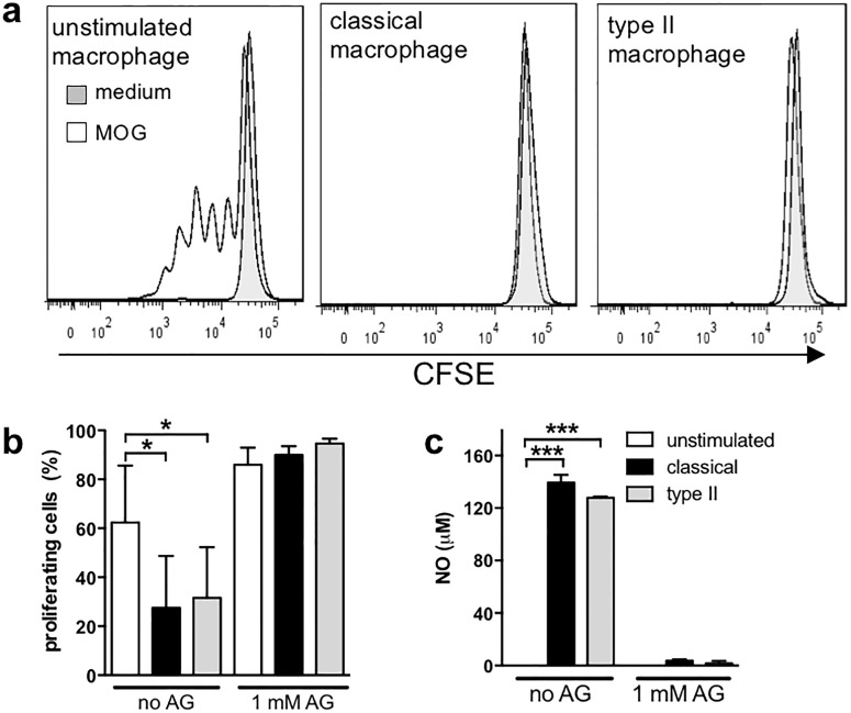
Fig 3. Type II macrophages do not enhance proliferation of CD4+ T cells in the absence of NO.
Macrophages were stimulated as described in Fig 2 in the presence or absence of 1 mM aminoguanidine (AG). After four hours, purified CFSE-labeled CD4+ 2D2 T cells and MOG (25 μg/ml) were added to the cultures and cultured for 72 hours. Proliferation was analysed by flow cytometry, CD4+ T cells were gated as shown in Fig 2 and CFSE positive cells were gated as indicated using CFSE unlabeled cells in Fig 2. (a) Representative plots of CFSE staining from the different treatment conditions are shown (Solid line with MOG, filled line without MOG). (b) Percentage of cells that proliferated was calculated. (c) NO levels were measured by Griess reaction. Shown are the means and SEM combined from 3 independent experiments (b) or a representative experiment of 3 (c). *p<0.05 and ***p<0.001 by one-way ANOVA with Neuwman-Keuls’ post-test.

