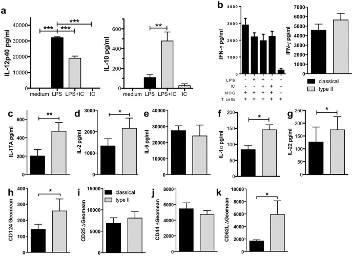
Fig 6. Type II microglia biased CD4+ T cell responses to a mixed Th17/Th2 phenotype.
(a) Microglia can be type II-activated. Microglia were isolated from the CNS of adult mice (n = 5/experiment) and expanded in the presence of M-CSF (5 ng/ml) for 4 weeks. Microglia were primed with IFN-γ (20 U/ml) overnight before stimulation with LPS (200 ng/ml) with or without IC (106/well) for 24 hours. Shown are the means and SEM of triplicate wells from a representative experiment of 3 independent experiments. **p<0.01 and ***p<0.001 by one-way ANOVA with Newman-Keuls’ post-test. (b-k) Type II microglia enhanced IL-17A production by CD4+ T cells. Microglia were isolated and activated as described. After four hours, purified CD4+ 2D2 T cells and MOG35-55 (25 μg/ml) were added for 72 hours. IFN-γ (b), IL-17A (c), IL-2 (d), and IL-6 (e) levels were measured by ELISA and IL-1α (f) and IL-22 (g) levels by CBA. CD124 (h), CD25 (i), CD44 (j), and CD62L (k) were assessed by flow cytometry. Shown are the means 7 (b-d, h-k) or 3 (e & g) independent experiments or the means and SEM of triplicate wells from a representative experiment of 3 independent experiments (b & f). *p<0.05, **p<0.01, and ***p<0.001 by one-way ANOVA with Newman-Keuls’ post-test (a) or Wilcoxon matched pairs signed rank test (b-k).

