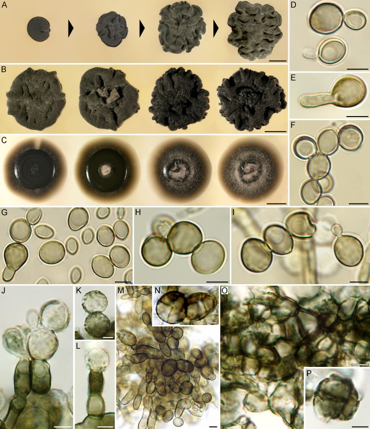
Fig 8. Macromorphology and micromorphology of Bacillicladium lobatum.
(A) Morphogenesis of colony growing on MEA at 25°C in 9 d, 2, 4 and 6 wks (left to right). (B, C) Phenotypic variability of 6-week-old colonies at 25°C on PDA (B) and PCA (C). (D‒I) Yeast-like state, budding (D, I), germinating by hyphae (E, G) or forming short chains (F‒I). (J‒L) Fungal elements from the inner parts of the colony with incrustations on their surface, occasionally proliferating. (M, N) Uni- or multicellular bodies, single or in chains. (O) Meristematic parenchyma-like structures formed in the inner parts of the colony (MEA). (P) Multicellular element released from the parenchymatous structure with roughened wall. Bar = 5 mm (A‒C), 5 μm (D‒P).

