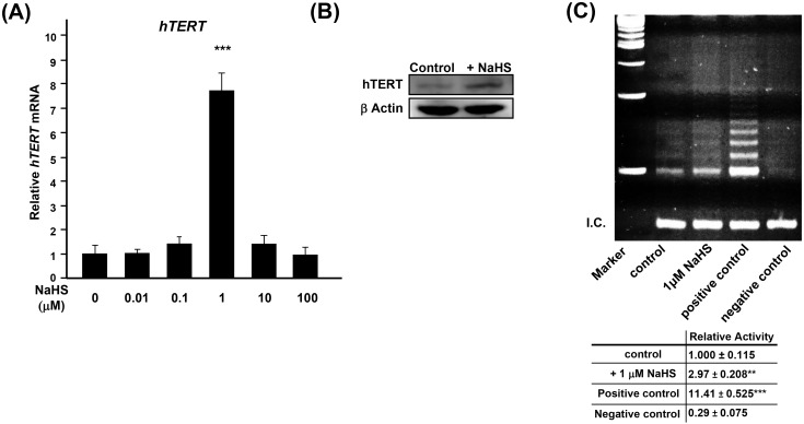
Fig 2. Exogenous H2S increases the expression of hTERT as well as the activity of telomerase.
(A) Real-time PCR analysis of the expression of hTERT in young (PD: 5.9) aHDF cells, treated with NaHS for 3 days. The expression of hTERT was normalized to the level of expression of β-ACTIN. Expression of untreated control was regarded as 1.0. (B) Immunoblotting of hTERT in aHDF cells without or with 1 μM NaHS for 7 days. 100 μg of the indicated nuclear extracts were subjected for immunoblotting. β-Actin was used as a loading control. (C) Telomerase activity in young (PD: 3.2) aHDF cells without or with treated with 1 μM NaHS for 7 days. Positive control was MDA-MB-231 cell lysate, and negative control was buffer alone. Bottom panel shows quantified means ± error bars from three independent assays. Relative activity of telomerase was calculated by dividing the density of all ladders to the density of the bands in internal control, indicated as internal control (I.C.).

