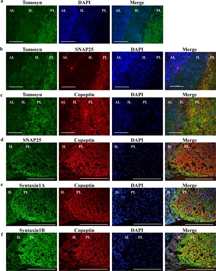
Fig 2. Tomosyn localizes in posterior pituitary.
(a) The cryosections of rat posterior pituitary were single stained with anti-tomosyn or DAPI (labelled on the top). The merged image is show at the right. (b–f) Double stained images are shown and the antibodies used are shown on top each image. Merged images are shown at right. Scale bars indicate 100 μm (white bars). AL, anterior lobe of the pituitary; IL, intermediate lobe; and PL, posterior lobe.

