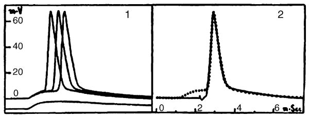
Fig. 2.
A reproduction of Figs. 1 and 2 in Brock et al. (1951a). These are the first known illustrations of an intracellular record from a vertebrate central neuron. The original figure legend read: “Figs. 1 and 2. Intracellular action potentials of motoneurons as described in the text. Potential and time scales common to both figures.” (p. 15). Their Fig. 1 illustrates orthodromically evoked APs evoked by stimulation of group I afferents, with the EPSP shown below. Their Fig. 2 illustrates an antidromically evoked AP, with the dotted line demonstrating a superimposed orthodromic spike. Reprinted with permission of the publisher.

