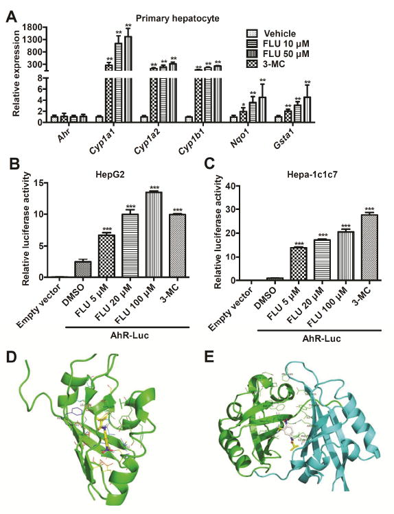
Fig. 3.

FLU stimulates mice AhR battery gene transcription. (A) qPCR analysis for AhR battery gene mRNA expression in mouse primary hepatocytes after FLU exposure. Significance was determined by two-tailed Student’s t-test test (*P < 0.05, **P < 0.01, versus vehicle group). (B and C) Luciferase assays for AhR activation in HepG2 (B) and Hepa-1c1c7 (C) cells. ***P < 0.001, compared with that of AhR-Luci+DMSO, by two-tailed Student’s t-test test. (D and E) Docking orientation of FLU into mouse AhR-LBD (PDB id: 4GHI) (D) and AhR dimer (PDB id: 4EQ1) binding pocket (ICM v3.5-1n, Molsoft) (E). The protein backbone is displayed as ribbon and colored by secondary structure. The residues are displayed as sticks and colored by atom type, with the carbon atoms in green. The ligands are displayed as sticks, colored by atom type, with carbon atoms in yellow or white, oxygen atom in red, fluorine atom in cyan, and nitrogen atom in blue.
