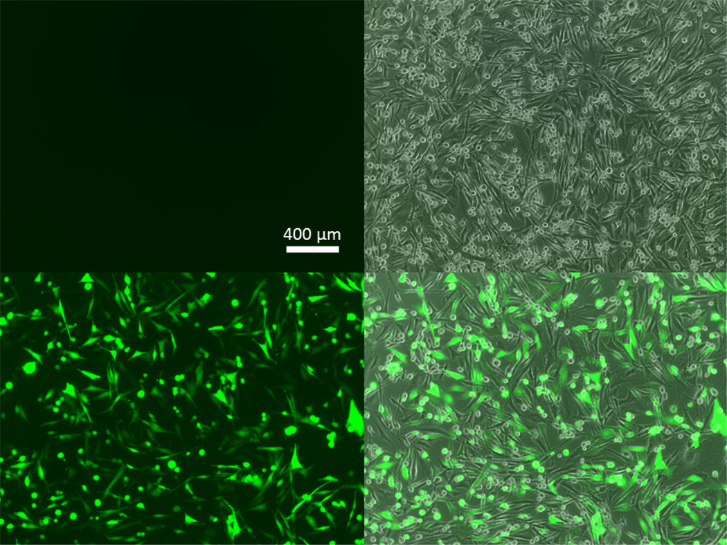
Figure 4. GFP transfection of human brain cancer cells with PBAE 447 polyplexes.
Fluorescence microscopy images of human primary glioblastoma cells showing the eGFP channel (left) and the eGFP and phase contrast channels combined (right) for untreated cells (top) and for 447 30 w/w polyplex transfected cells (bottom) at 48 hours post transfection.

