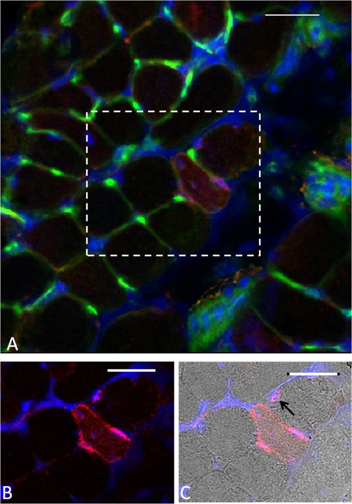
Fig. 3.
a Example of WGA–fluorescein lectin staining (green) and NCAM immunostaining (red) on a transverse cross-section of an EDL muscle from an old mouse at 3 days following damaging lengthening contractions. DAPI staining (blue) was used to visualise the nuclei. b Focused image of the denervated fibre. c Bright field image of the denervated fibre. Black arrow indicates satellite cell. Scale bar 30 μm

