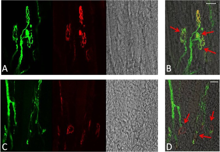
Fig. 6.
Representative longitudinal sections of EDL muscles from quiescent old mice showing YFP in motor axons (green), AChRs labelled with Alexa-594-α-bungorotoxin (red) and image of the fibres under bright field (a, c). b, d Merged image. Red arrows show age-related structural changes including nerve terminal fragmentation with some nerve terminals forming spherical and partially or fully denervated NMJs. Scale bar 30 μm

