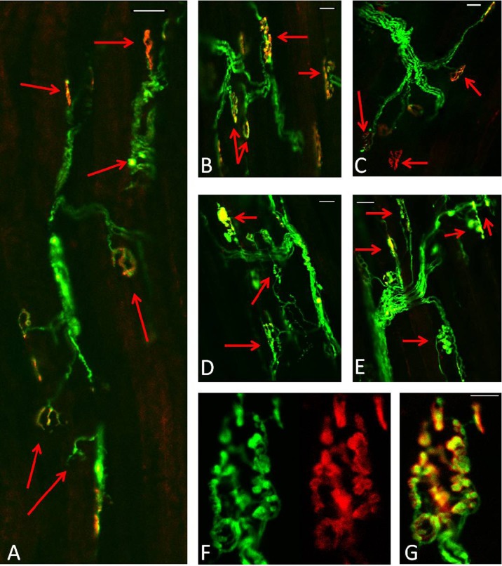
Fig. 7.
Longitudinal sections of EDL muscles from old mice expressing YFP in motor axons (green). a Three days following damage, b–c 28 days post damage, d–e 60 days following damage. AChRs labelled with Alexa-594-BTX (red). Scale bar 30 μm. f–g Higher magnification of a NMJ from EDL muscle of an old mouse 60 days following damage. AChRs labelled with Alexa-594-BTX (red). Scale bar 10 μm

