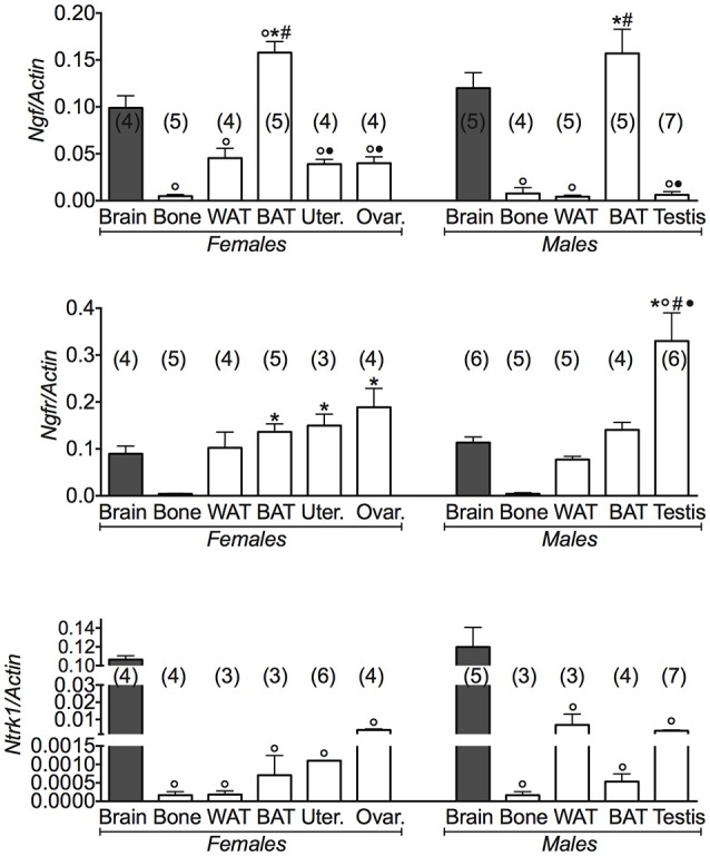
Figure 3.

NGF gene and its receptors in different tissues of 3 months old female and male mice. Relative expression levels of NGF gene and its receptors in brain, bone, white adipose tissue (WAT) and brown adipose tissue (BAT), uterus, ovaries, testis of 3 months old female and male mice. The genes under investigations were: nerve growth factor Ngf and associated receptor nerve growth factor receptor Ngfr, and neurotrophic tyrosine kinase, receptor, type 1 Ntrk1. Brain data were reported for comparison as a positive control. In each graph, the bars are the means ± SEM from the n animals indicated in brackets. For the Ntrk1 gene, 2 out of 5 analyzed samples in WAT and BAT from female mice, and 4 out of 7 analyzed samples in bone and WAT from male mice were not amplified due to the extremely low expression levels of this gene in these tissues. Differences were found within groups and between groups as determined by one way analysis of variance for Ngf, Ngfr, and Ntrk1. Data significantly different (P < 0.05) vs. brain (◦), bone (*), WAT (#), and BAT (•) using Bonferroni analysis.
