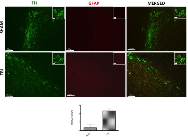
Figure 4.
Effect of chronic TBI on Tyrosine hydroxylase (TH) expression in glial fibrillary acidic protein (GFAP) positive cells. Midbrain tissues were double-stained with antibodies against GFAP (red) and TH (green). The red spots indicate the co-localizations (merged) indicated by yellow arrows. Midbrain sections revealed increased astrogliosis (GFAP+ cells) in TBI panels. TH positive neurons were significantly reduced after TBI (TBI merged panel). Scale bar = 50 and 20 μm (particle). Each data are expressed as Mean ± SEM from N = 5 mice/group. Counting of colocalized cell confirmed our data. The co-localization of image was analyzed with image J software.

