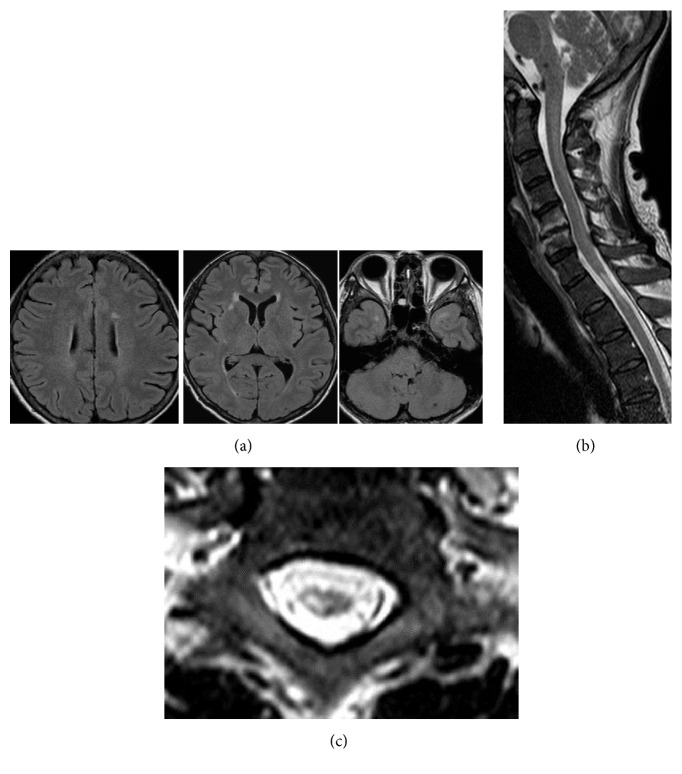
Figure 1.
Characteristic magnetic resonance images (MRI) of Case 1. Cranial MRI showed small white matter and subcortical lesions suggestive of intracranial HTLV-I-associated myelopathy lesions (a). On spinal cord MRI examinations, a marked high signal intensity lesion was observed in the thoracic spinal cord at the level of the T1 and T2 segments (b). The spinal cord showed regional atrophy, and a T2 high-signal lesion was found mainly in the right central part of the spinal cord (c).

