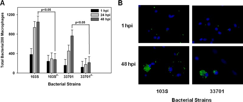
FIG 3 .
Equine R. equi isolates containing the pVAPA1037 plasmids replicate in swine macrophages. The intracellular growth of equine R. equi strains 103S, 103SP−, 33701, and 33701P− (A and B) was assessed in swine monocyte-derived macrophages infected at an MOI of 5:1. Triplicate monolayers were fixed and stained at 1 h, 24 h, and 48 h postinfection (hpi), and the number of bacteria per 200 macrophages was determined (A). Representative microscopy images of monolayers infected with R. equi strains 103S and 33701 at 1 h and 48 h postinfection are shown (×60 magnification) (B). In these images, R. equi exhibits green fluorescence and the macrophage nucleus is blue because of DAPI staining. Statistical analysis was performed on the data compiled from 2 individual experiments.

