
Figure 4.
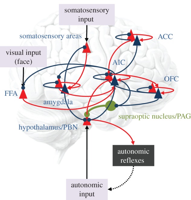
A (simplified) neural architecture underlying the predictive coding of visual, somatosensory and interoceptive signals. The anatomical designations, although plausible, are used to simply illustrate how predictive coding can be mapped onto neuronal systems. As in figure 2, red triangles correspond to neuronal populations (superficial pyramidal cells) encoding prediction error, while blue triangles represent populations (deep pyramidal cells) encoding expectations. These provide descending predictions to prediction error populations in lower hierarchical levels (blue connections). The prediction error populations then reciprocate ascending prediction errors to adjust the expectations (red connections). Arrows denote excitatory connections, while circles denote inhibitory effects (mediated by inhibitory interneurons). In this example, recurrent connections mediate innate (epigenetically specified) reflexes—such as the suckling reflex—that elicit autonomic (e.g. vasovagal) reflexes in response to appropriate somatosensory input. These reflexes depend upon high-level representations predicting both the somatosensory input and interoceptive consequences. The representations are activated by somatosensory prediction errors and send interoceptive predictions to the hypothalamic area—to elicit interoceptive prediction errors that are resolved in the periphery by autonomic reflexes. Oxytocin (in green) is shown to project to the hypothalamic area, to modulate the gain or precision of interoceptive prediction error units. One hypothesis for autism rests on a failure to attenuate the precision of autonomic prediction errors, thereby precluding expectations about visual and somatosensory information (e.g. a mother's face or affiliative touch) that is not accompanied by autonomic input (see the text). FFA, fusiform face area; AIC, anterior insular cortex; ACC, anterior cingulate cortex; OFC, orbitofrontal cortex; PAG, periaqueductal grey; PBN, parabrachial nucleus. (Online version in colour.)