Figure 1.
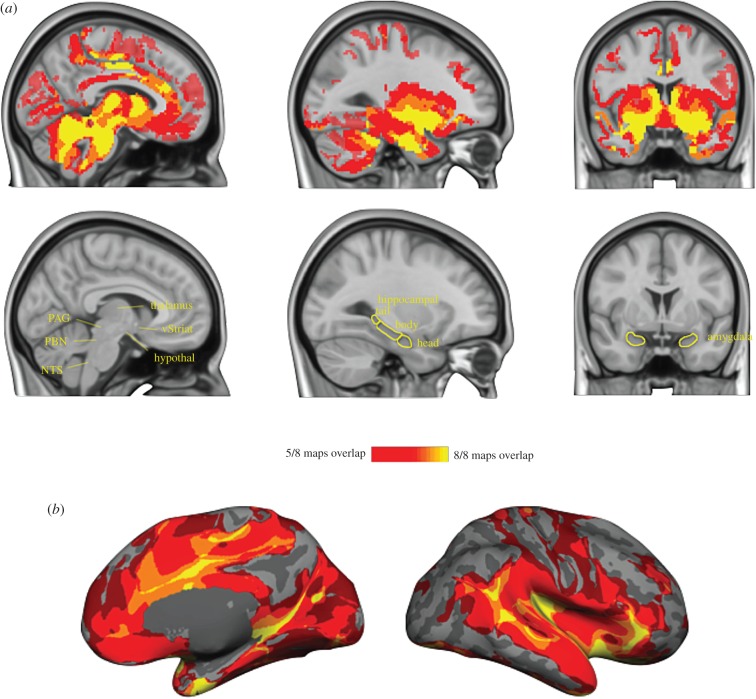
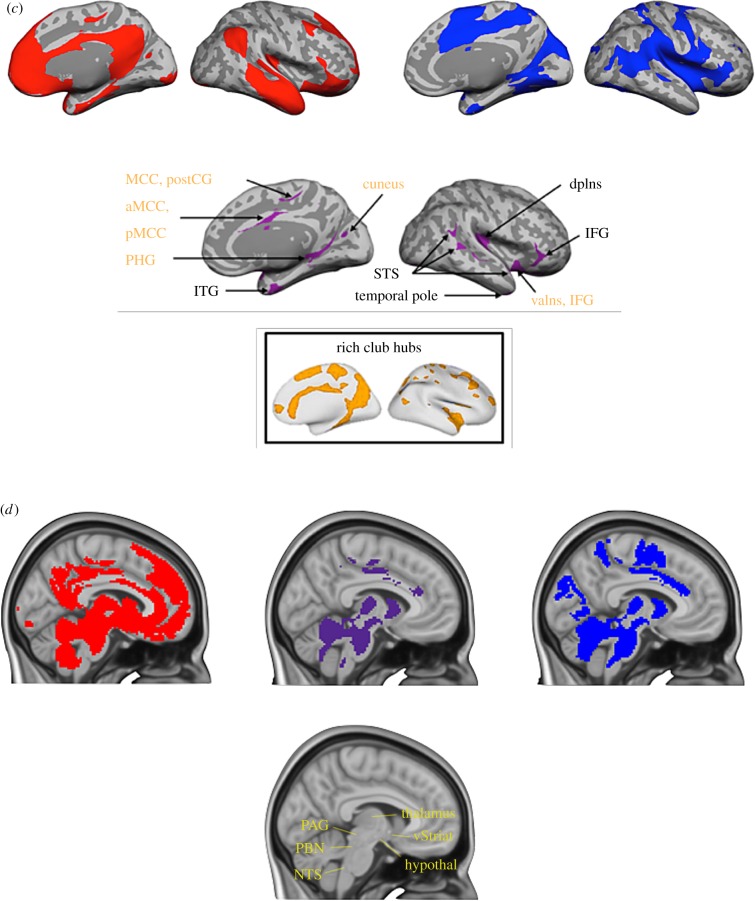
A large-scale system for allostasis in the human brain. We consulted anterograde and retrograde tract-tracing studies in macaque monkeys to select eight seed regions in limbic cortices with monosynaptic connections to midbrain and brainstem regions that are known to control the immune, endocrine and autonomic nervous systems in the service of allostasis (for details and coordinates, see [52]). For each seed region, we computed a ‘discovery map’ of voxels whose timecourse correlated with the seed region. (a) A conjunction of all eight maps presented in the volume to display subcortical regions. (b) A conjunction of maps depicted on the cortical surface. (c) Cluster analysis of the eight discovery maps revealed the system for allostasis was composed of two large-scale intrinsic networks (shown in red and blue) that share several hubs (shown in purple). Hubs belonging to the brain's ‘rich club’ are labelled in yellow. Rich club hubs figure adapted with permission from [54]. Maps were constructed with resting state BOLD data from 280 participants binarized at p < 10−5, and then replicated on a second sample of 270 participants. aMCC, anterior midcingulate cortex; dpIns, dorsal posterior insula; IFG, inferior frontal gyrus; ITG, inferior temporal gyrus; vaIns, ventral anterior insula; MCC, midcingulate cortex; PHG, parahippocampal gyrus; pMCC, posterior midcingulate cortex; PostCG, postcentral gyrus; STS, superior temporal sulcus. (d) Reliable subcortical connections, thresholded p < 0.05 uncorrected. PAG, periaqueductal grey; hypothal, hypothalamus; PBN, parabrachial nucleus; vStriat, ventral striatum; NTS, nucleus of the solitary tract.


