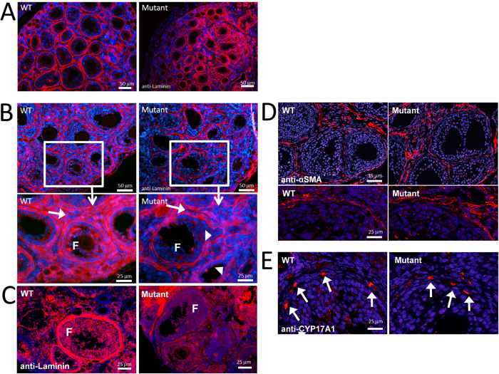
Figure 6. Loss of GAS2 expression disrupts organization of the basal lamina during follicular development.
Anti-laminin (red) immunofluorescence labeling of the basement membrane with cell nuclei counterstaining by DAPI (blue) highlights the ECM structure surrounding follicles in the P7.5 wild type and mutant mice (A). The basal lamina of the Gas2 null mutant mice was disorganized compared to wild type mice. At P12.5 this disorganization appeared more severe (B). Higher magnification highlights the stromal compartment between growing follicles (arrows), which has less dense and organized laminin staining. Arrowheads point to impaired integrity of the basal lamina in Gas2 null ovaries compared to wild type. Laminin staining of adult ovaries (C) showed that the basal lamina surrounding growing follicles remains poorly organized into adulthood in Gas2 null mutant females; however this does not appear to be due to a reduction in laminin protein expression (D), as determined by Western blot analyses (D), 1 = ovary, 2 = kidney). Staining of α-SMA (E) and CYP17A1 (F) demonstrated that vascular structures and thecal cell differentiation appear normal in the Gas2 null mutant mice.

