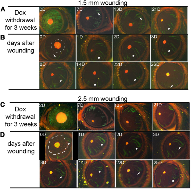
Figure 3. Response of limbal epithelial progenitor to corneal epithelial wounds.
The Krt12R/TC/Rosa26F/GFP triple mice at five weeks of age were injected with 80 μg/g body weight Dox in 0.9% aqueous NaCl (10 mg/ml stock) followed by Dox-chow (1 g Dox/kg chow) for 4 days, then fed with regular chow. The corneal epithelial injury was performed at 3 weeks after Dox withdrawal to ensure that only progenitor derived green strips were stabilized and observed. The green strip appearance and changes before and after wounding were recorded by photography. (A) Photographic images of the same eye at different days after Dox withdrawal. (B) Photographic images of the same eye at different days after 1.5 mm cornea epithelium debridement performed at 3 weeks after Dox withdrawal. The wound areas are outlined by white dashed lines. The white arrows point to a same green strip over the time. (C,D) Photographic images of another eye underwent a similar procedure as above, but wounded with 2.5 mm cornea epithelium debridement. The images shown are representatives from 5–6 eyes examined for each condition.

