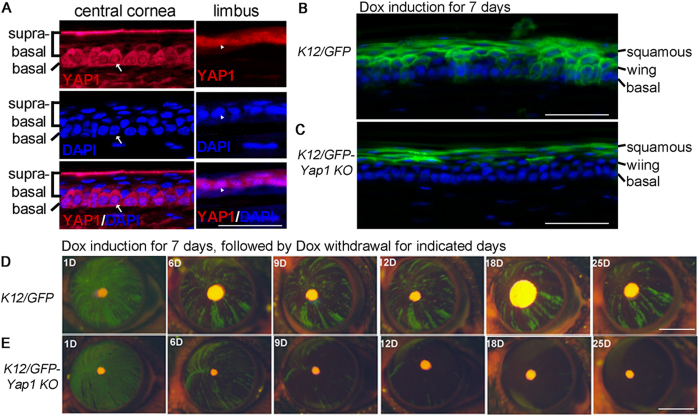
Figure 4. Requirement of YAP1 for maintenance of the cornea epithelial progenitors in vivo.
(A) Nuclear YAP1 was detected in the limbal and corneal basal cells. DAPI was used for nuclear counterstaining. The arrows and arrowheads indicate the same position in the corneal and limbal epithelia, respectively. (B,C) Mosaic GFP expression at cornea basal layer of Krt12R/R/TC/Rosa26F/GFP (K12/GFP) mouse at 7 week old (B), but absent in the age matched Krt12R/R/TC/Rosa26F/GFP/Yap1fl/fl (K12/GFP-Yap1KO) mouse (C). The images were taken immediately after 7 days of Dox treatment. (D,E) Live images taken from a Krt12R/R/TC/Rosa26F/GFP (K12/GFP) mouse (D) and Krt12R/R/TC/Rosa26F/GFP/Yap1fl/fl (K12/GFP-Yap1KO) mouse (E) at the different time points after Dox withdrawal. The mice at five weeks of age were fed with Dox diet for 7 days, then followed by regular chow without Dox for indicated days. Scale bars = 35 μm in (A), 50 μm in (B,C), 2 mm in (D,E). The images shown are representatives from 5 eyes examined.

