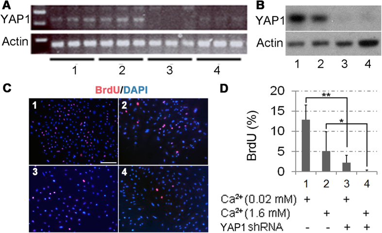
Figure 5. YAP1 regulation of the corneal epithelial cell proliferation in vitro.
Primary mouse corneal epithelial cells were transduced with YAP1-specific (sample 3 and 4) or scramble control (sample 1 and 2) shRNA lentiviruses for 2 days; and then replaced with fresh medium containing 0.05 (sample 1 and 3) or 1.6 mM (sample 2 and 4) calcium and purimycin for additional 48 h. Data are representatives of three independent experiments. (A) RT-PCR showed that YAP1 was knocked down in primary corneal epithelial cells. (B) Loss of YAP1 expression in the shRNA knockdown cells was further confirmed by Western blot. (C) The representative images of BrdU staining (red) on the primary mouse cornea epithelial cells. Cells were pulse-labeled with BrdU for 90 min to label the proliferating cells. DAPI (blue) was used for nuclear counterstaining. (D) Quantitation of BrdU-positive cells. Data are expressed as mean ± SD. Statistical significance was analyzed by two-tailed Mann-Whitney U test: *P < 0.05, **P < 0.005. Scale bar = 150 μm in (C). The images shown were representatives from 3 independent experiments.

