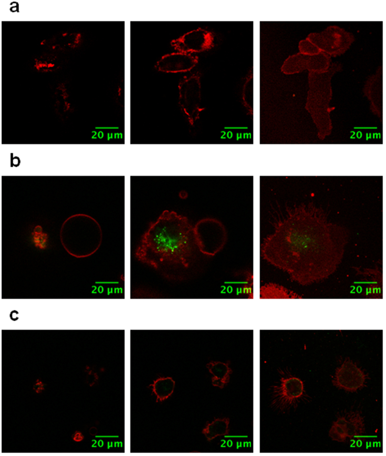
Figure 3. Polyman26 pulse-chase internalization experiments.
Optical slices of iMDDCs incubated with 50 μM Polyman26 (pseudo-colored in green) in HBSS and, after 3 washes with HBSS (150 μL), stained for 2 min at RT with DiD’ (pseudo-colored in red) as membrane marker. After the last washing steps (2 washes with 150 μL of HBSS), cells were fixed with 1% PFA. Left: dorsal membrane (i.e. exposed to the media); centre: middle of the cell body; right: ventral membrane (i.e. in contact with the culture plate). (a) Negative control; (b) iMDDCs were incubated with 50 μM of Polyman26 at 37 °C for 60 min; (c) iDCs were incubated with 50 μM of Polyman26 at 4 °C for 20 min.

