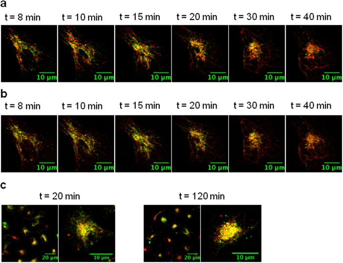
Figure 4. Subcellular localization of Polyman26 after internalization.
Images were taken with a confocal microscope on live cells, at the time intervals indicated in the panels. Polyman26 is pseudo-colored in green and the tracers are pseudo-colored in red. (a) iMDDCs were incubated with Polyman26 (50 μM) plus OVA 488 in HBSS, for 10 min at RT and analysed after a washing step at indicated time points. (b) iMDDCs were incubated with Polyman26 (50 μM) plus Transferrin 633 in HBSS, for 10 min at RT and analysed after a washing step at indicated time points. (c) iMDDCs were incubated with 100 μM Polyman26 in HBSS for 20 min at RT (left) or 120 min at 37 °C (right); after a washing step, LysoTracker was added and cells were analysed.

