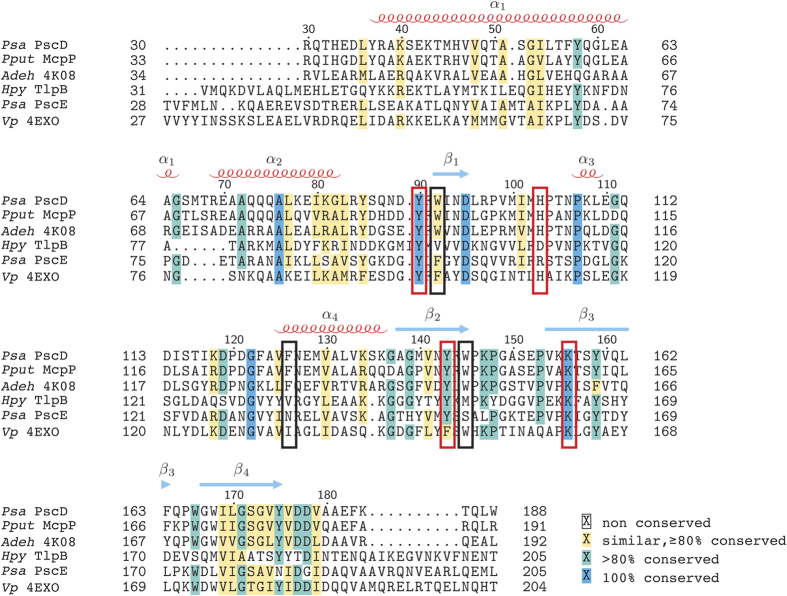
Figure 5. Multiple sequence alignment of PscD-SD with other known single Cache chemoreceptor sensor domains.
The alignment was performed using MUSCLE with shading in TEXShade by LATEX 2ε using shading mode similar. Sequences are labelled by species abbreviation and either protein name or PDB code. Secondary structural elements of PscD-SD are mapped onto the alignment with alpha-helices (labelled α1-α4) indicated by red coils and beta-strands (labelled β1-β4) indicated by blue arrows. Red boxes indicate amino acids that form polar contacts with the ligands and black boxes indicate other amino acids in the binding-site.

