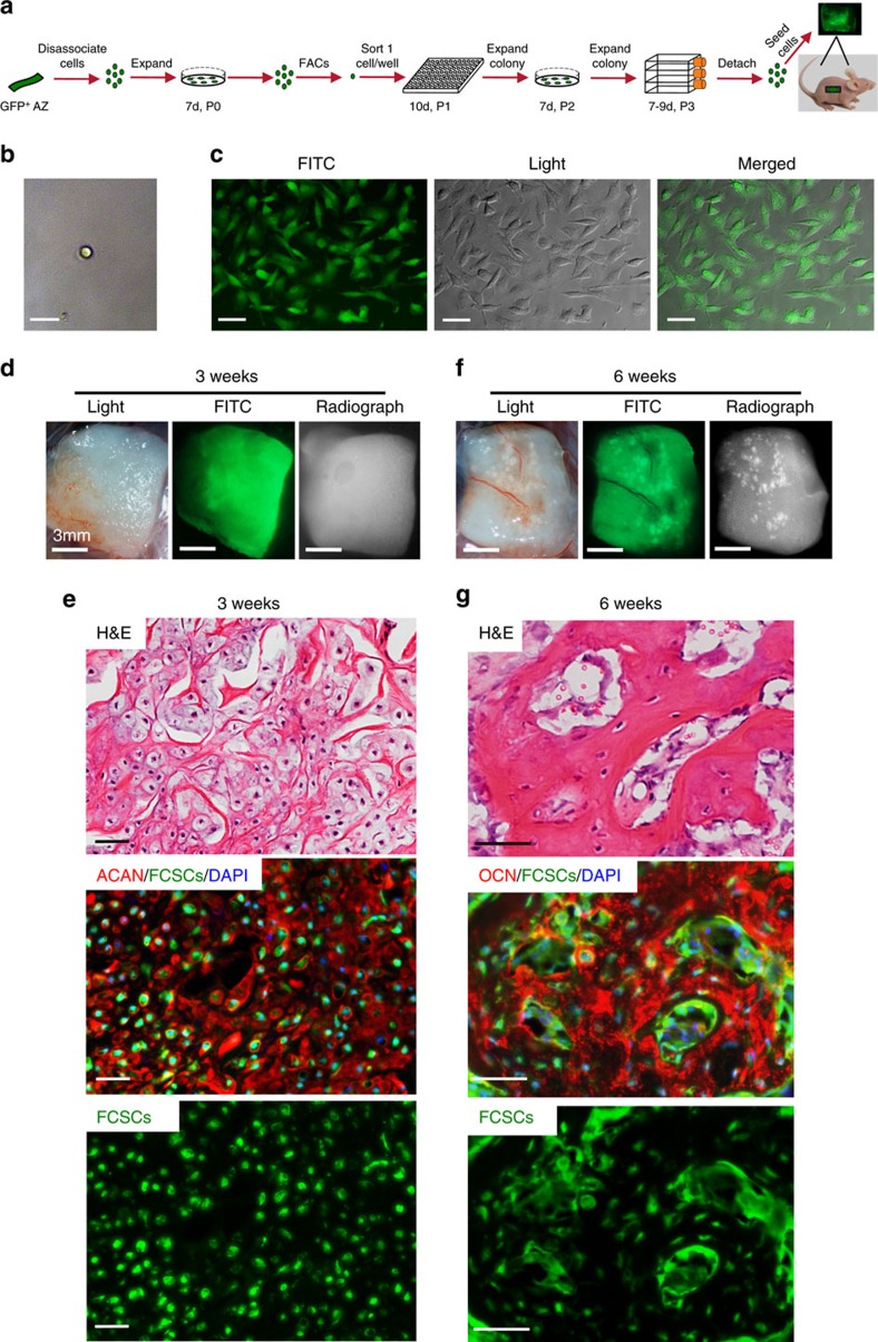
Figure 3. A single FCSC spontaneously generates cartilage and organized bone.
(a) Schematic representation showing single FCSC isolation. Heterogeneous GFP+ FCSCs were derived from the TMJ SZ tissue from male GFP transgenic rats 8 weeks. GFP+ FCSCs were expanded in vitro and FACs was used to plate a single-cell per well into a 96-well plate. Each single-cell colony was expanded over passages 2–3, seeded onto a collagen sponge and surgically transplanted subcutaneously on dorsum of nude mice. (b) Sorted single GFP+ FCSC in a single-well/96-well plate. Scale bar, 25 μm (c) GFP+ FCSC single-cell colony expanded at P3. Scale bar, 50 μm. (d,f) Collagen sponge seeded with GFP+ FCSC single-cell colony on the dorsum of nude mice after 3 and 6 weeks in vivo, respectively. Scale bar, 50 μm. (e,g) Representative H&E staining GFP+ FCSC transplant from a single-cell after 3 and 6 weeks, respectively (top row). Immunohistochemistry for aggrecan (ACAN, red) and osteocalcin (OCN, red) demonstrated single-cell GFP+ FCSC generated cartilage at 3 w and bone at 6 w, respectively (middle and bottom rows). Dapi blue staining=nuclei. H&E, haematoxylin and eosin.

