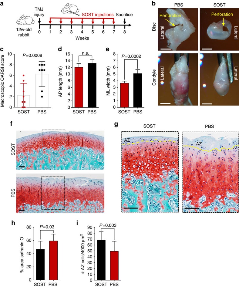
Figure 5. Sclerostin induces endogenous FCSCs to regenerate and repair cartilage.
(a) To induce fibrocartilage injury and condyle degeneration, a 2.5 mm perforation was created in 12-week rabbit TMJ discs bilaterally. One week post surgery, 0.1 ml SOST (100 ng ml−1) and PBS was injected into contralateral sides of the inferior TMJ intra-articular space once per week for 7 weeks. (b) Superior view of TMJ disc and condyle 8 weeks following injury (yellow arrow) with SOST and PBS treated contralateral TMJs. Scale bar, 3 mm. (c) Observers (n=2) blindly scored TMJ morphology using OARSI recommendations. Scatter plots with each plot representing mean rabbit score±s.d., n=8 male New Zealand White rabbits 12 weeks; paired Student's t-test. (d,e) TMJ condyle anterior to posterior length (AP) and the medial to lateral (ML) width was measured using Nis-Elements Basic Research imaging, respectively. Data are mean; error bars are s.d.; n=8 male New Zealand White rabbits 12 weeks; paired Student's t-test. (f,g) Representative images of Safranin O staining of contralateral TMJ condyles treated with SOST and PBS. Yellow line=border of SZ tissue. Scale bar, 100 μm. (h) Safranin O staining in TMJ condyle was quantified using cellSense Imaging Software. Data are mean area±s.d.; n=8 male New Zealand White rabbits 12 weeks; paired Student's t-test. (i) The number of FCSCs quantified in SZ tissue. Data are mean±s.d.; n=8 male New Zealand White rabbits 12 weeks; paired Student's t-test.

