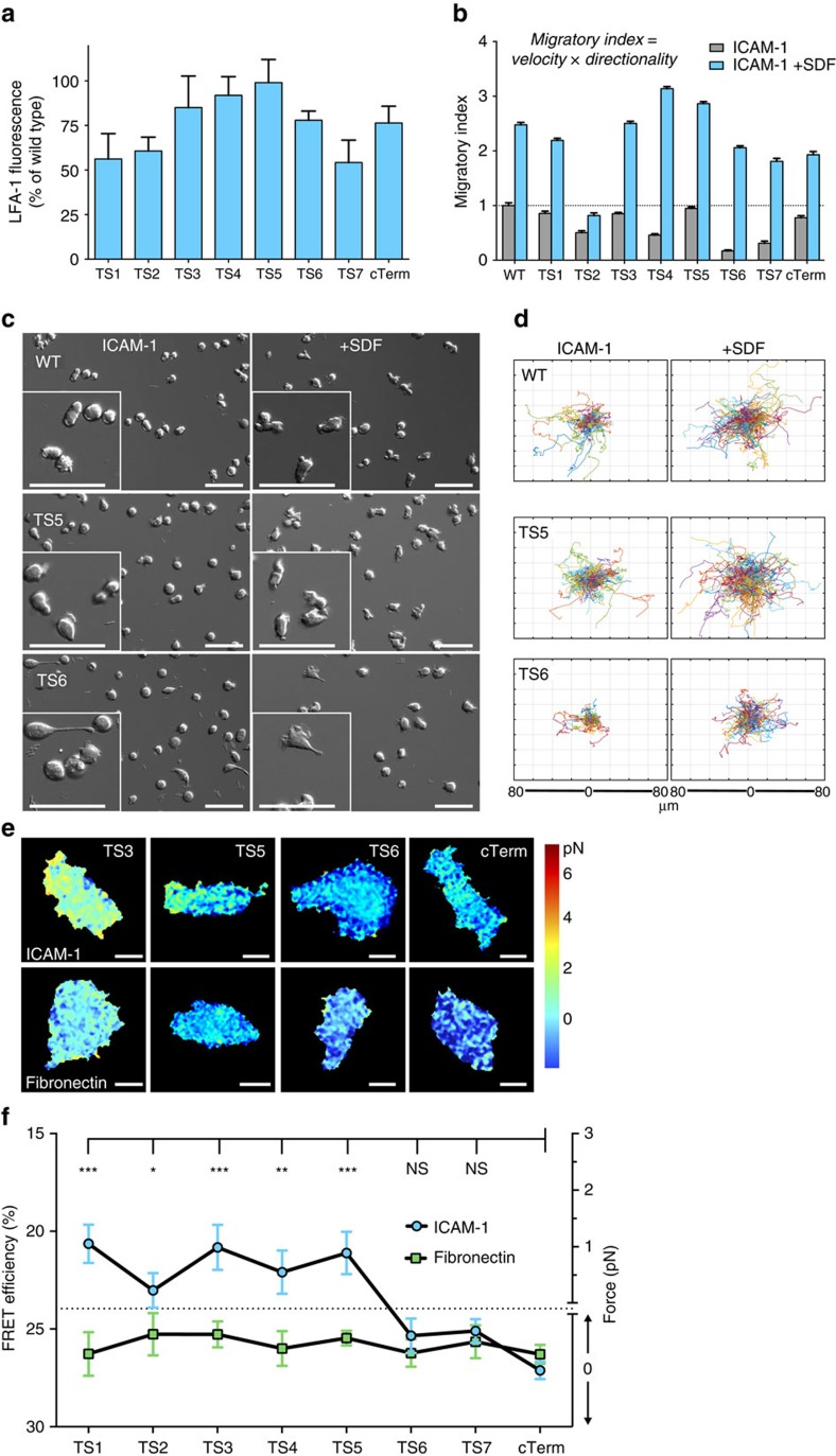
Figure 2. β2 tension sensors placed N-terminal to actin adaptor-binding sites respond to force.
(a) Transient expression of β2 tension sensors in 293T cells. Each sensor was transfected together with wild type αL. LFA-1 was quantitated by flow cytometry with the heterodimer-specific β2 monoclonal antibody TS1/18. Average values±s.e.m. from three independent transfections. (b) Migration of Jurkat cells on 20 μg ml−1 ICAM-1 with or without 100 ng ml−1 SDF-1α. Each β2 construct was introduced by lentiviral transduction without addition of αL to allow pairing with wild type αL. WT is non-transduced Jurkat cells. From left to right: N=166/187; 177/293; 102/121; 355/317; 215/350; 315/409; 186/233; 169/164; 348/159. Average values with±s.e.m. are shown. Data are from three independent experiments. (c) Representative time-lapse DIC images of cells used for quantification of (b). Scale bars, 50 μm. (d) Representative cell tracks of cells used in (b) with all starting points shifted to the center. (e) Representative FRET images from fixed Jurkat T cells. Scale bar, 5 μm. Images are from 4 to 5 independent experiments. (f) Quantification of whole-cell FRET levels in fixed Jurkat T cells that were migrating on either ICAM-1 or fibronectin. Values are shown as mean +−s.e.m. Kruskal–Wallis with Dunn's multiple comparison test for β2-cTerm on ICAM-1 and the others yielded P-values: ***P<0.001, **P<0.01, *P<0.05. No significant differences found when comparing cells on fibronectin.

