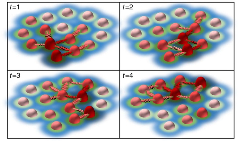
Figure 9. Schematic representation of the dynamic spatial patterns within podosome clusters.
Individual podosomes (light to dark red cones) continuously undergo growth and shrinkage cycles. Within a cluster, podosome neighbours are connected by myosin decorated actin filaments (red arrows) that drive a spatial coordination of actin and force oscillations resulting in a wave-like phenomenon (t=1 to t=4, arbitrary time units). These continuous actin waves directly drive the recruitment of vinculin (green) and talin (blue) resulting in mesoscale dynamic spatial patterns within the cluster that extend well beyond the nearest neighbour. For simplicity, the actin filaments connecting individual podosomes to the cell membrane are not depicted.

