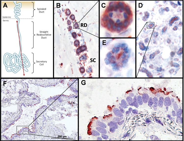
Figure 3.
Immunohistochemical staining of CA XII and CA II in human skin and lung. (A) Diagram depicting longitudinal view of sweat gland components with red line indicating a hypothetical plane used to generate slices for the micrographs shown in (B)–(E). (B) Two populations of sweat gland resorptive ducts (‘RD’) and secretory coils (‘SC’) immunostained for CA XII and counter-stained with hematoxylin and eosin. The resorptive ducts (n = 9 different cross-sections captured in this panel) show positive staining for CA XII in a two cell thick layer of cuboidal epithelia cells. The myoepithelial cells surrounding the secretory coils (n = 3 different cross-sections captured in this panel) show light staining of CA XII that may be non-specific. Magnification of this micrograph is 100×. (C) Enlargement of CA XII positive staining of the basolateral membrane in resorptive ductal cells from (B). (D) Positive staining of apically localized control protein CA II in sweat ducts and secretory coils. Magnification of this micrograph is 100×. (E) Enlargement of resorptive ducts from (E) showing apical localization of CA II. (F) CA XII positive staining of the luminal edge of a bronchiole cross-section. Magnification of this micrograph is 100×. (G) Positive staining of apically localized CA XII in the terminal bar of bronchial epithelia. Magnification of this micrograph is 400×.

