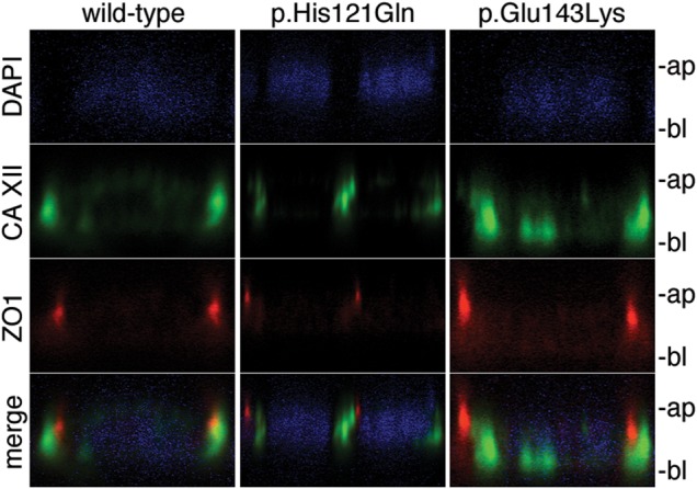
Figure 6.

Subcellular localization of WT and mutant CA XII in polarized MDCK cells. Fluorescent co-staining of (left) WT CA XII (green), (center) p.His121Gln (green), and (right) p.Glu143Lys (green) with endogenous tight junction protein ZO1 (red) and nuclear stain DAPI (blue) in polarized MDCK cells imaged in the xz-plane. This micrograph reveals primarily lateral staining of CA XII; however, basal and lateral staining were observed for WT (n = 7 different micrographs), p.His121Gln (n = 8 different micrographs), and p.Glu143Lys (n = 13 different micrographs). The apical membrane is indicated by ‘ap’ and the basal membrane is indicated by ‘bl’.
