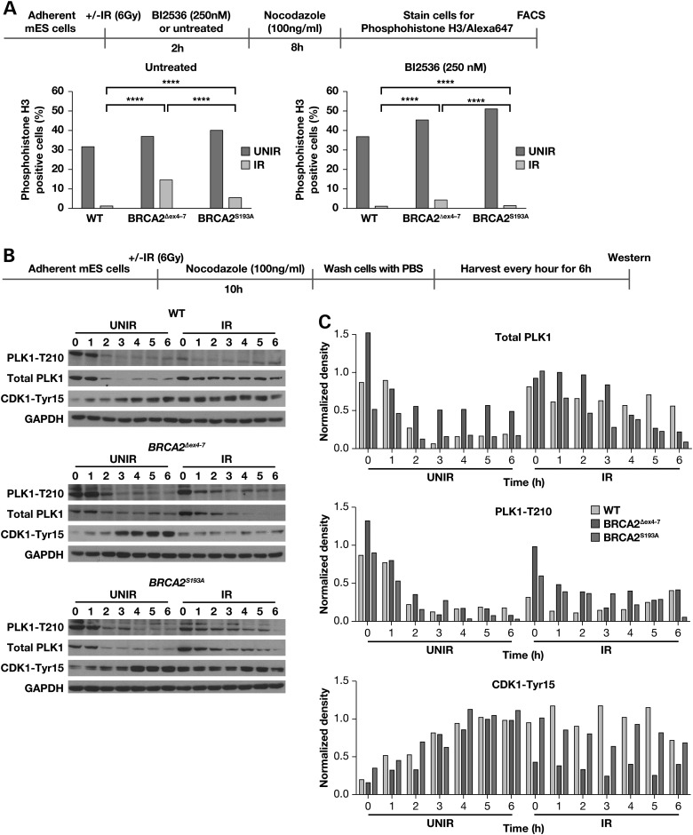
Figure 5.
Aberrant activation of PLK1 following IR in BRCA2Δex4–7 expressing mES cells. (A) FACS analysis of phospho-histone H3 (Ser10) stained mES cells expressing WT, BRCA2Δex4–7 and BRCA2S193A with or without PLK1 inhibitor treatment, following IR. (B) Western blot analysis of total cell lysate from mES cells expressing WT, BRCA2Δex4–7 and BRCA2S193A showing total PLK1, activated PLK1-Thr210 and CDK1-Tyr15 levels at different time following release from nocodazole. (C) Densitometric analysis of protein bands shown in (B), normalized against glyceraldehyde-3-phosphate dehydrogenase. The scheme of experimental design is shown at the top of each panel.

