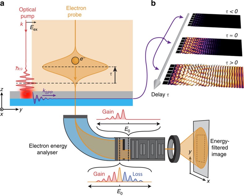
Figure 1. Time-resolved PINEM methodology.
(a) Simplified scheme of the time-resolved photon-induced near-field electron microscopy (PINEM) experiments in this work. A photon pump pulse incident on a nanopatterned Ag-on-Si3N4 bilayer structure generates a surface plasmon polariton (SPP) wave propagating along the buried Ag/Si3N4 interface. The near-field of the propagating SPP is subsequently probed through its interaction with a field-of-view electron pulse at a time delay τ. Energy-filtered imaging of the resulting electron distribution of transmitted electrons then provides spatially resolved temporal snapshots of the near-field corresponding to the propagating plasmonic wave. (b) Variation of the relative time delay between the optical excitation pulse and the probing electron pulse generates a time-resolved movie of the ultrafast evolution of the buried plasmonic near-field.

